Cell Animation (Metastasis)
Our Process: Movements involved in cancer cell formation
In an assignment from the Department of Biomedicine at the University of Baselwe were faced with a challenge of visualizing amoeboid cell movements involved in the formation of cancer cells. The so-called epithelial-mesenchymal transition (EMT) in the the initiation of cancer mechanisms and is subject of foundational research.
The goal of the project was to show the dislocation of cells as well as the structures of the cytoskeleton involved in the movement in two short animated clips.
Client
University of Basel
Department of Biomedicine
Client
University of Basel
Department of Biomedicine
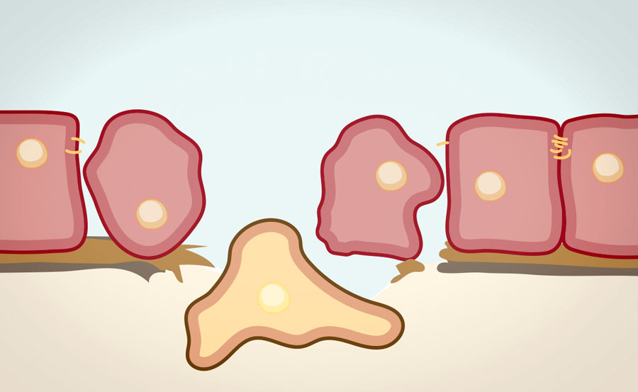
Animation of Amoeboid Cells
As in a typical scientific diagram, the representation of the cells and their situation should be schematic and simplified. This does however not imply any lack of inaccuracy: Structures, and – in this specific case – amoeboid cell movements, had to be rendered in a natural and credible way.
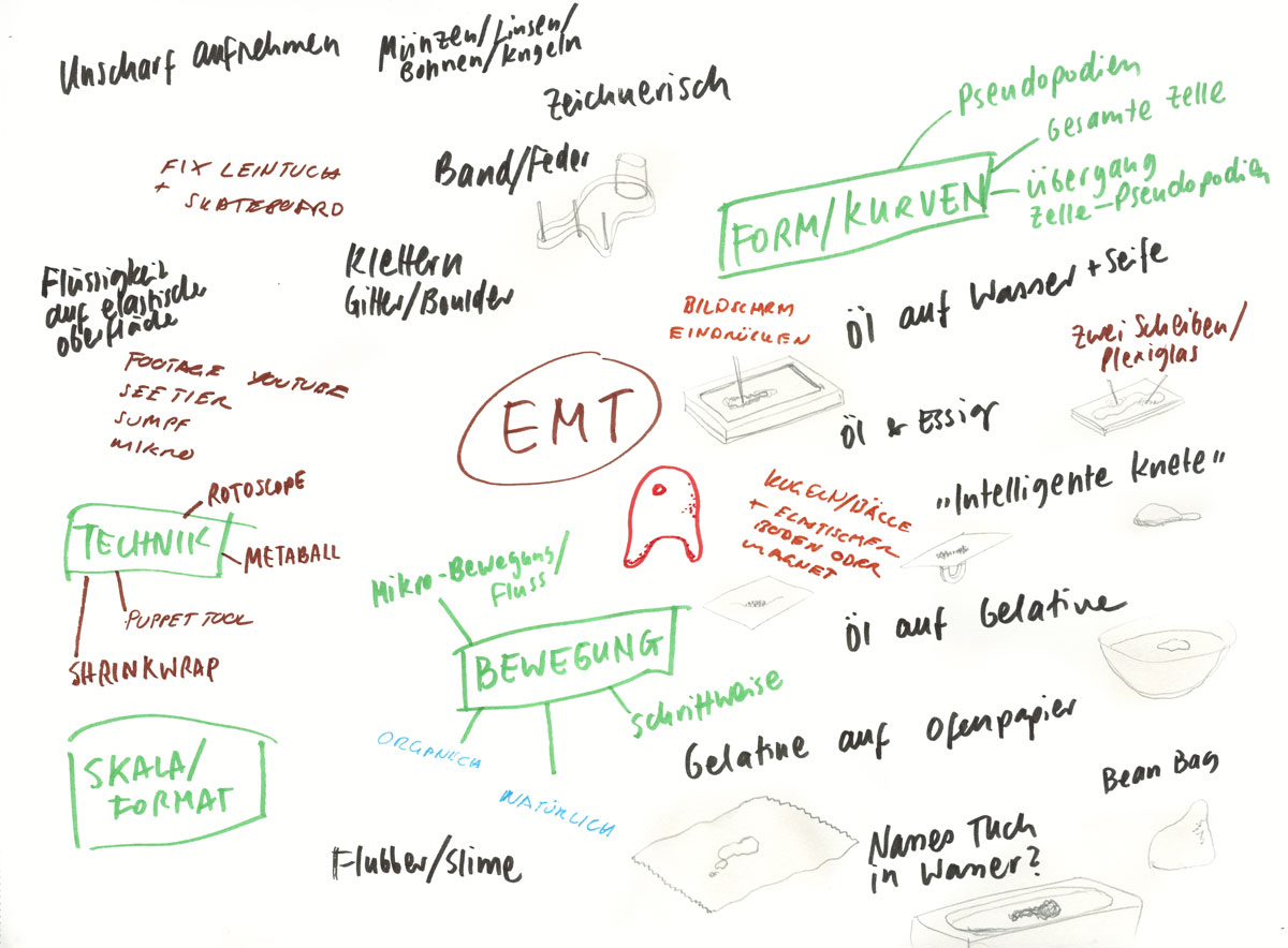
Experiments with fluid media
In search of a way to simplify the flowing amoeboid movement while still maintaining an organic appeal, we conducted various experiments with fluids. Oil on water, ink on water-repellent paper and boiling drops of water (leidenfrost effect) were some ideas to recreate the moving blob-like structure. An analogy to the invasive nature of mesenchymal cells also can be found in swarm movement.
First Results
After editing the videos created in the experiments, the outlines of the blobs could be directly traced to create an animated 3D mesh of the cells. As the footage was mostly too short to serve as a basis for the whole movement, we had to freely interpret the subsequent deformations. This gave us a significant freedom in designing and timing the animation while supporting us with a solid foundation of ‘realistic’ movement.
Visualization of Cytoskeletal Structures
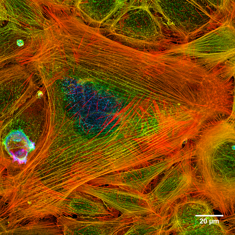
The cytoskeleton is a complex network of filaments and tubules which acts as a scaffolding for the cell’s shape, is involved in cell signaling pathways, uptake and secretion of materials, and can actively contract in order to make the cell move. In this assignment we were asked to visualize the cytoskeletal structures involved in the EMT. Image: Actin filaments within a cell, visualized by confocal fluorescence microscopy. CC-BY-SA Howard Vindin (Wikipedia)
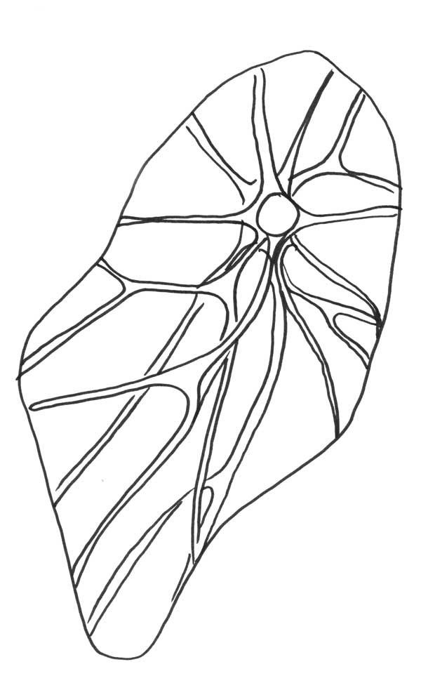
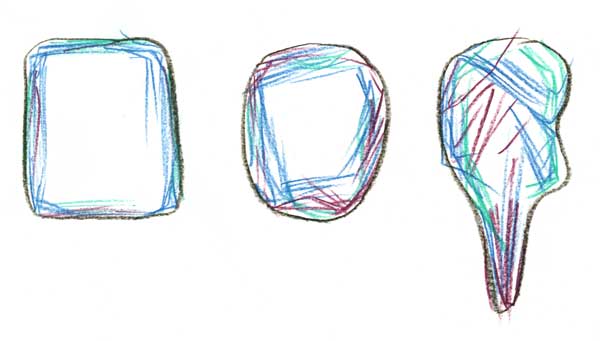
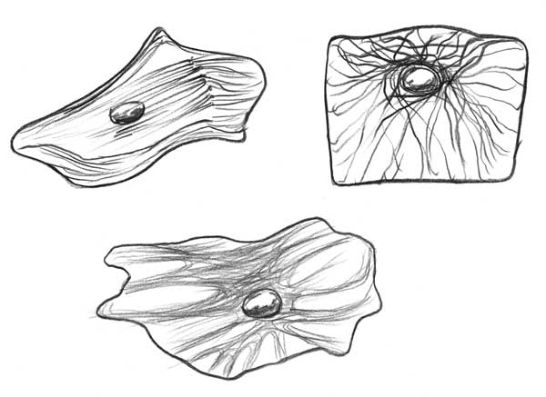
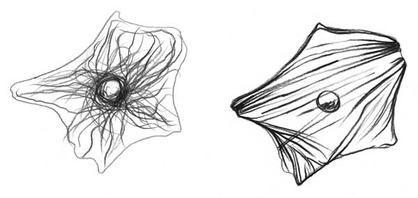
Insight by sketching
The cytoskeleton is composed of various structures that serve different functions. It was mainly through sketching that we understood that it would make sense to only show one cytoskeletal component, namely the microfilaments. Consisting of actin filaments, the microfilaments are tense polymers that generate force pushing against the cell membrane and thus are involved in the movement of the cell. Other components of the cytoskeleton are intermediate filaments and microtubules, which are involved in junctions and intracellular transport. The most important visual difference (relevant for our animatiion) is that they look more loose than the actin microfilaments and create a visible structure around the nucleus.
Results
As in microscopy the cytoskeleton is visualized by fluorescence, we chose to imitate this aesthetic to make the structures recognizable as such at first glance. The animation acts therefore as an overlay based on the first clip, where only the cells’ silhouettes and nuclei are visible.